How Core Needle Biopsy Improves Diagnosis of Musculoskeletal Tumours: Insights from a Singapore-Based Research
Accurate diagnosis is a critical step in the management of musculoskeletal tumours, which include a wide range of bone and soft tissue lesions. Tissue sampling plays an essential role in distinguishing between benign and malignant conditions, guiding subsequent clinical decisions. Core needle biopsy has emerged as a frequently used diagnostic approach in orthopaedic practice. A Singapore-based research study involving Dr Seng Chusheng and colleagues has evaluated the diagnostic performance of core needle biopsy for musculoskeletal tumours, providing insights relevant to clinicians and patients alike.
Understanding the Challenges of Diagnosing Musculoskeletal Tumours
Musculoskeletal tumours encompass both bone and soft tissue lesions that may be benign, malignant, or inflammatory in nature. Distinguishing between these categories is necessary for planning appropriate management, whether surgical or non-surgical. Clinical examination and imaging studies such as radiography and magnetic resonance imaging often provide initial information, but definitive diagnosis generally requires histological confirmation from a tissue sample.
Open surgical biopsy has long been considered the conventional standard for tissue diagnosis. However, it can involve a surgical procedure with associated recovery, costs and risk of complications. As a result, there has been clinical interest in less invasive techniques that can obtain representative tissue samples with fewer risks.
What Is Core Needle Biopsy in Orthopaedic Practice?
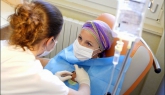
Core needle biopsy is a minimally invasive procedure in which a hollow needle is used to extract cylindrical tissue samples from a lesion for histopathological analysis. In the orthopaedic setting, this procedure may be performed with imaging guidance—such as ultrasound, computed tomography (CT) or other modalities—to improve accuracy in targeting the lesion. The technique allows the clinician to obtain multiple cores of tissue under local anaesthesia, often in an outpatient setting.
The aim of core needle biopsy is to obtain sufficient tissue to enable pathologists to distinguish between benign and malignant tumours and to provide subtype information where possible. Its use complements clinical assessment and imaging in establishing a diagnosis while potentially reducing the need for more invasive surgical biopsy.
Overview of the Singapore-Based Research Study
The research published in the Journal of Orthopaedic Surgery (Hong Kong) by Chusheng Seng and colleagues evaluated the diagnostic performance of core needle biopsy for musculoskeletal tumours at a Singapore institution. This study retrospectively reviewed records of patients who underwent core needle biopsy followed by definitive surgical excision, comparing the biopsy results with the final histological diagnosis obtained after surgery.
The primary objective was to assess how closely the initial core needle biopsy results matched the definitive diagnoses, focusing on sensitivity and specificity in differentiating benign and malignant lesions. Analysis included a range of tumour types affecting bone and soft tissues.
Key Findings on Diagnostic Accuracy
In the study cohort of 134 patients who underwent both core needle biopsy and subsequent surgical excision, the findings showed a high level of concordance between core needle biopsy results and final histology. For 118 of the tumours (88 per cent), the biopsy results matched the final diagnosis. Assuming that the small number of non-diagnostic cases would have been correctly classified, the calculated sensitivity was 95 per cent and specificity was 97 per cent in differentiating benign versus malignant lesions.
A small proportion of cases yielded non-diagnostic results due to insufficient tissue sampling, and in a minority of cases there were mismatches between biopsy findings and final histology, including instances where the lesion was initially classified as benign but subsequently found to be malignant. These outcomes highlight both the strengths and limitations of the biopsy technique under real-world clinical conditions.
Clinical Implications for Orthopaedic Care
The results of this study suggest that core needle biopsy, when performed with appropriate technique and sufficient tissue sampling, can provide a highly concordant diagnosis with final surgical histology for many musculoskeletal tumours. This has practical implications for orthopaedic practice, where accurate preoperative diagnosis influences decisions about surgical planning, oncological management and multidisciplinary care.
Clinicians may consider core needle biopsy as part of the diagnostic work-up for musculoskeletal lesions, recognising that it aims to reduce diagnostic uncertainty and may inform the need for further investigation or referral to specialist services.
About the Research Contributor
Dr Seng Chusheng holds a Bachelor of Medicine and Bachelor of Surgery and a Master of Medicine in Orthopaedic Surgery from the National University of Singapore. He is a Member and Fellow of the Royal College of Surgeons of Edinburgh and has completed subspeciality training in foot, ankle and lower limb conditions in Geneva, Switzerland.
Concluding Perspective
Accurate tissue diagnosis remains fundamental in the evaluation of musculoskeletal tumours. Core needle biopsy offers a diagnostic option that may complement other clinical and imaging findings, potentially reducing the need for more invasive procedures in selected cases. Research such as that conducted by Seng and colleagues contributes to the understanding of diagnostic tools in orthopaedic practice, supporting informed clinical decision-making.
References
- Accuracy of core needle biopsy for musculoskeletal tumours. Journal of Orthopaedic Surgery (Hong Kong), Seng C et al., 2013. Sensitivity and specificity of core needle biopsy were 95% and 97% respectively in a retrospective series of 134 patients.
- Best in Singapore. (n.d.). Orthopaedic specialist profile: Dr Seng Chusheng. Retrieved from https://www.bestinsingapore.co/best-orthopaedic-specialist-singapore/